Digital Thermography
What is Thermography?
It is a non-invasive Infrared Thermal Imaging with
No Radiation and
No Compression
Easy Preventative Screening
Being proactive with your health and well-being is the best way to live a long, productive life. Practicing prevention is paramount. There are many ways you can support your body’s long term health, and an excellent first step is knowledge of the “current state of affairs” regarding your body’s functioning.
Current research has shown that inflammation is the foundation of most modern diseases. Tests like MRI, X-ray, mammogram, and ultrasound are all tests of structure and are not capable of imaging inflammation so that it can be addressed early.
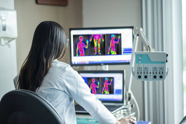
Imagine having a tool that aids your preventive healthcare efforts by revealing inflammation in your body, potentially altering outcomes. Enter Digital Infrared Thermal Imaging (DITI), the sole screening tool capable of visualizing inflammation and associated heat patterns.
This non-invasive, painless imaging technology detects abnormal blood flow and inflammatory indicators by capturing the invisible infrared heat emitted by your body. These images highlight areas of inflammation, serving as early warning signs for various abnormalities, conditions, syndromes, and diseases.
Armed with this information, you and your healthcare provider can formulate a plan to address any issues detected for a long, healthy life.

Thermography Services
Breast Screening
Baseline Screening
Every woman has a unique thermal heat pattern. It is as unique as her fingerprint. Because thermal imaging looks for change over time, it is essential that a stable baseline be established. This requires two sets of images be taken within a 3-4 month period. This helps to assure that the breast tissue is unchanged and remains stable. Once a baseline is established, it will then be used for comparison to all future scans so that the most subtle of tissue changes can be identified.
Routine Screening
The standard recommendation is that all women should begin annual breast screening at age 40. With recent statistics indicating that 1 in 8 women will be diagnosed with breast cancer during their lifetime, early detection is critical.
Urgent Screening
Early detection is critical and can make the difference between a complete recovery and a shortened lifetime. If you have a breast concern and need urgent screening, we will make you a priority.
Full-Body Screening
Can be useful for the evaluation of:
- Early indications of asymptomatic inflammatory/degenerative processes
- Systemic inflammation
- Unexplained Pain
- Referred Pain
- Arthritis
- Fibromyalgia
- Digestive Disorders
- Artery Inflammation
- Nerve damage
Head and Neck Screening
Can be useful for the evaluation of:
- TMJ Dysfunction
- Dental Inflammation
- Carotid artery inflammation
- Thyroid over/under active
- Cervical spine evaluation
- Sinus inflammation
- Trigeminal Neuralgia
- Referred pain
- Headache evaluation
Upper Body Screening
Can be useful for the evaluation of:
- TMJ Dysfunction
- Dental Inflammation
- Carotid artery inflammation
- Thyroid over/under active
- Cervical spine evaluation
- Sinus inflammation
- Trigeminal Neuralgia
- Referred pain
- Headache evaluation
Chest Screening
Can be useful for the evaluation of:
- Shoulder pain
- Inflammation
- Esophageal/GERD inflammation
- Brachial plexus injury
- Male breast cancer
Abdomen Screening
Can be useful for the evaluation of:
- Systemic inflammation
- Digestive disorders
- Unexplained pain
Back Screening
Can be useful for the evaluation of:
- Disc Disease
- Herniated Discs
- Myofascial Irritation
- Nerve Entrapment/Impingement
- Referred Pain Syndrome
- Trigger Points
Arm Screening
Can be useful for the evaluation of:
- Shoulder pain
- Inflammation
- Carpal Tunnel Syndrome
- Tendonitis
- Nerve entrapment
- Sprain/Strain
- Peripheral Nerve injury
- Raynaud’s Syndrome
Leg Screening
Can be useful for the evaluation of:
- Inflammation
- Deep Vein Thrombosis
- Neuropathy
- Ligament tear
- Tendonitis
- Nerve entrapment
- Sprain/Strain
- Peripheral Nerve injury
Imaging can help locate the problem
Conditions and Symptoms that can be explored with Thermal Imaging

Breast Cancer concerns and early Detection
Thermography measures the heat coming from your body. Metastatic cancers create heat which can be imaged by digital infrared imaging. As thermal imaging has been demonstrated in numerous studies to be capable of measuring these heat signatures years before conventional technologies can see a mass, and as the procedure uses no radiation, compression of breast tissue and as it is totally safe, thermography or DITI provides for a safe early warning detection system.
Arterial Conditions
The ability of thermography to detect the presence of deep vein thrombosis and other circulatory disorders of the lower extremities is a very exciting application of this procedure as it allows us to painlessly and safely detect possible disease that if unchecked, could cause the loss of a limb, or in some cases add to the possibility of stroke. Another aspect of thermal imaging which has gone largely unnoticed is in developing diabetic neuropathies of the feet, before the foot becomes insensate.
Musculoskeletal and Sports Medicine
This is one of the clearest examples of thermography's ability to accurately diagnose patients with a host of back, neck and extremity disorders. When muscle tissue is strained or torn, it releases chemicals which cause increased heat. This can be seen as intense patterns of hyperthermia in the region of the muscle, or trigger point, as in the case of fibromyalgia.
Nerve Damage
Nerve damage, as occurs in disc herniation and spinal nerve root compression, displays on the thermographic map in exactly the opposite direction as muscle injury by revealing cool areas of hypothermia in the nerve tracts coming from the spine. This also makes it possible to detect Diabetic Neuropathy as well.
Screening for Arthrits
Thermography indicates where thermal temperature changes, so for inflammatory arthritis, the local temperature increases due to increased vascularity in the inflamed tissue area.
Understanding Thermography
Thermal Imaging has so many beneficial uses
Watch this short video to get a better understanding of how you or a loved one may benefit from Thermal Imaging.
Completely Non-invasive
There is no radiation, compression, or even touching of your body.
Tailored to You
Thermal Imaging is always customized to each individual based on their concerns, symptoms and history.
Affordable
We have worked hard to be able to offer our patients very affordable fees and efficient service.
Frequently Asked Questions
What is DITI?
Digital infrared thermal imaging, or DITI, is a totally non-invasive, radiation-free, painless procedure with no contact with the body. DITI is a clinical imaging procedure that records the patterns of infrared heat naturally emitted from your body. Your thermal images can be used by your healthcare practitioner to help assess pathology in the body as well as monitor pain. Thermography is a test of vascular physiology, meaning that it looks for abnormal blood flow, inflammation and functional changes in the tissue which can often be the indicator of early-stage disease.
How does thermal imaging work?
Thermal imaging is a 15 minute, non-invasive test of vascular physiology. Thermal imaging uses a highly sensitive, high-resolution digital thermal camera to take a picture of your body’s infrared heat and display these patterns in the form of a digital image. The cells of your body produce heat through their normal function. Abnormal cells typically produce more heat in the early stages of development (before a tumor forms).
Before most tumors grow, abnormal cells will:
- stimulate new blood vessels to grow
- re-open unused blood vessels
- maintain those blood vessels already in use
The cells build a vast network of blood vessels in the area it will ultimately grow. A tumor needs more blood flow than normal cells to support its rapid growth. With more blood flow, there is more heat. Thermal imaging examines these “hot spots” which can be the early warning signs of developing pathology. This activity has been shown to begin years before a tumor forms, and before any warning signs can be given by other screening methods.
Is thermography safe?
The camera used by Hormones by Design for imaging is the only FDA registered thermal imaging medical camera on the market specifically designed for medical screenings. It is a totally non-invasive procedure that is completely safe. There is absolutely NO contact with the body, NO compression, and NO radiation. While a variety of studies have called into question the safety of cumulative exposures to radiation, this is not the case with thermography. Thermography emits nothing. It is simply a picture of your unique thermal heat patterns. It is a quick and painless procedure, which makes it a great screening option for breast screening with “no harm done” in the process.
Who reads the images and writes the reports?
Our images are sent to Electronic Medical Interpretation, Inc. EMI is a professional group of physicians who are trained in the protocols of reading thermal images. A formal interpretation and written report, including color images, is prepared and sent to you in approximately two weeks. If quicker results are desired, rush service is available for a fee. We are happy to email you a copy of your report so that you can share it with your other physicians upon request.
How accurate is thermography?
Thermography has a sensitivity and specificity rate of approximately 90%. As with any test, results are often only as good as the technician performing them. Don’t hesitate to ask how much experience your technician has with the equipment and the performance of the procedure. A positive thermogram can be the single most important marker of high risk for developing pathology.
Who can benefit from breast thermography?
Breast thermography is a great option for all women but particularly women that fall into certain categories. Women with dense breast tissue, fibrocystic breasts, as well as women with implants or women that have had mastectomies without reconstructive surgery are more difficult to screen for breast cancer using other screening technologies. However, that is not the case with thermography. Since the screening process is nothing more than taking pictures or images of the infrared heat emitted from the body, the size of the woman, size of the breasts or tissue type are no longer concerns for the sake of breast screening. This test can provide a clinical marker to the doctor or practitioner in the event that a specific area of the breast needs particularly close or frequent examination.
Why do I need to establish a baseline for breast thermography?
Every woman has a unique thermal heat pattern – it is as unique as their fingerprint. Because of this, it takes two sets of images to establish a baseline. The first set of images provides the first half of your baseline of your unique “thermal pattern”. Because thermal imaging looks for changes over time, it is essential that a second set of images be completed within 3-4 months after the first set of images.
This subsequent session assures that the thermal patterns remain unchanged. Once a stable baseline is established, it will be used for comparisons to all future scans so that the most subtle of tissue changes can be identified.
What is the difference between thermography and mammography?
Thermography detects the subtle physiologic changes that accompany breast pathology, whether it is cancer, fibrocystic disease, an infection, or vascular disease. It can alert you and your doctor to changes that can indicate early stage breast disease. It is safe, effective, and ideal for women of all ages. It is completely non-invasive and does NOT use radiation. It is a “do no harm” approach to routine breast screening.
Mammography is a test of anatomy that uses x-ray to look for masses or lumps. These masses can take years to form – sometimes 5-10 years before they are large enough and dense enough to show up on an x-ray. Although touted to be harmless, mammograms do involve compression of the breast tissue, which may be uncomfortable for some women. A dose of radiation is administered with each view taken.
While thermography is a great basic screening method, it looks at the breast tissue differently than a mammogram does. For some, thermography is an adjunctive procedure and is used in conjunction with the annual mammogram and/or ultrasound. For others who may not be candidates for mammography or choose not to use mammography, thermography is a great option. Mammography can be a very useful follow up tool when warranted. When used in conjunction with thermography, the rate of early detection is increased to as much as 95%.
In the event that something suspicious should appear on your thermogram, your doctor or health care practitioner should be consulted immediately. After a thorough discussion of your options, you and your doctor or health care practitioner may decide that a mammogram or ultrasound is warranted for a different view of the area of concern.
Can thermography diagnose breast cancer?
No. In fact, all other screening methods (mammography, ultrasound, physical/self-exam) can only suggest the presence or absence of disease. The only way to diagnose breast cancer is through a biopsy and pathological study.
What does the breast thermography procedure involve?
This quick and easy procedure starts with you undressing from the waist up and changing into a hospital gown to allow air to circulate which allows your body temperature to cool down. Cooling time is approximately 12 minutes. Once acclimated, you will be asked to sit in front of the camera, with your hands behind your head. Five different images will be taken. This allows the camera to scan the breasts, neck and underarms as well as the lymph nodes under the arms, breasts, and neck. These images are then sent for interpretation and archived for future comparison.
Is this procedure covered by insurance?
No. Payment for your scan is expected at the time of your screening. We accept cash, checks, MasterCard, Visa, Discover, and HSA and/or medical spending accounts. Please call our office for more specific details.
What is your cancellation policy?
If you are unable to keep your appointment, we ask that you show consideration by calling the appropriate clinic (Austin or San Antonio), one business day in advance of your scheduled appointment. Due to the distances we travel to our out of town clinics to provide you with this service, as well as staffing requirements and other pre-planned needs, we would like to have the option to offer your appointment to others who may not have been able to schedule an appointment due to lack of availability. If you fail to give us one business day notice, a $50 cancellation charge will be billed to your account.
Digital Thermal Imaging
Our Locations
Austin
2200 Park Bend Drive, Building 1, Suite 201
Austin, Texas 78758
Call to schedule an appointment:
737-258-4014
San Antonio
21218 Market Ridge, Suite 101
San Antonio, Texas 78258
Call to schedule an appointment:
210-305-5077

Testimonial
What They Say
I always feel welcomed, comfortable and safe when I go for my breast thermography with Michelle. And I like that I’m not radiating my breast every year. Overall it is a positive experience every time I go. Kathy who makes the appointments and runs the office is also very helpful and clarifying any questions I have had about my appointments, the procedure, and protocol. I highly recommend Diti Imaging for your wellness checks.


I have been going to DITI Imaging for more than 5 years for my breast Thermography. I do not like radiation on my breasts and lymph nodes so I choose thermography only. The staff are welcoming and very attentive to your needs. They are professional and kind. I highly recommend DITI for the exams you need. They send a copy of the exam to my gynecologist so the doctor is learning the benefits of breast thermography for women.
